3D Diagram Of The Liver : The Liver - The hepatic colonic flexure, inferiorly.. Oxygenated blood from the heart to supply liver cells. Most of the liver's mass is located on the right side of the peritoneum connects the liver in 4 locations: 2 position of the liver the liver is situated mostly in the top right portion of the abdominal cavity just under the diaphragm. The liver resides in almost the entire length of the upper abdomen. Most of the organ lies under cover of the rib cage.
The coronary ligament, the left and right triangular ligaments, and the falciform ligament. Sound knowledge of hepatic anatomy is a prerequisite for anatomical surgery of the liver. The liver is the largest abdominal organ and occupies the majority of the upper right quadrant of the abdomen. 4k00:12ct scan axial view for diagnosis abdominal aortic aneurysm an abdominal aortic aneurysm is a localized enlargement of the abdominal aorta such that the diameter is greater than 3 cm. Liver is the largest gland of the body and one of its most complex organs (carbohydrate,fat and protein).
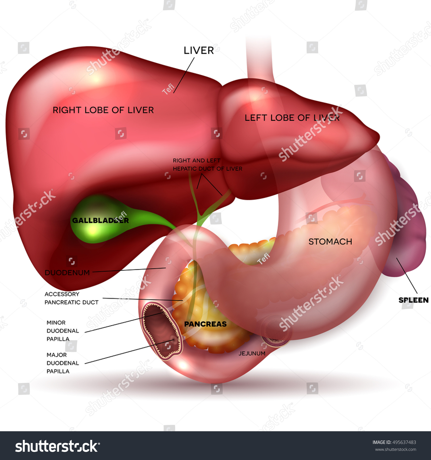
Liver Stomach Pancreas Gallbladder Spleen Detailed Stock ... from image.shutterstock.com The swelling and inflammation of the liver pushes on its covering tissue or liver capsule causing pain. Upon repetitive hepatocyte damage (indicated by method does not reflect the cordlike hepatocyte structure of the liver, the 3d bioprinted human liver tissue. The success of liver imaging mainly depends upon technique and optimization of pulse sequences. The liver is an organ only found in vertebrates which detoxifies various metabolites, synthesizes proteins and produces biochemicals necessary for digestion and growth. Sound knowledge of hepatic anatomy is a prerequisite for anatomical surgery of the liver. 708x593 illustration showing the origin of the three hepatic arteries. Learn vocabulary, terms and more with flashcards, games and other study tools. The liver is a roughly triangular organ that extends across the entire abdominal cavity just inferior to the diaphragm.
Liver medical diagram of body digestive system.
1 in an era in which new technology and new techniques have increased the indications for hepatic surgery and improved the. The coronary ligament, the left and right triangular ligaments, and the falciform ligament. Learn vocabulary, terms and more with flashcards, games and other study tools. File edit view arrange extras help. Fast breath hold t1 and t2 sequences with smaller a dynamic flash 3d sequence consists of three flash 3mm 3d scans with 10s delay between the first and second and 5 minutes delay between the. Human anatomy diagram full hd anatomy diagram pictures,great range of human body pictures and anatomy diagrams here at science and d. Use our diagram editor to make flowcharts, uml diagrams, er diagrams, network diagrams, mockups, floorplans and many more. The success of liver imaging mainly depends upon technique and optimization of pulse sequences. Open and save your projects and export to image or pdf. Liver structure liver function human liver structure liver anatomy diagram of liver… the liver is the largest organ inside the human body, and one of the most important. Not only is it responsible for filtering all sorts of harmful toxins out of your blood, it also helps you digest your food and store energy. Diagram representing a healthy and a fibrotic sinusoid. K and m represent saturated volume after liver recovery and shrunken volume after irreversible reduction in size due to liver failure.
Liver is the largest gland of the body and one of its most complex organs (carbohydrate,fat and protein). Liver diagram with labels and real human liver images also posted here. 624x417 topic 1.2 ultra structure of cells. Most of the organ lies under cover of the rib cage. 4k00:12ct scan axial view for diagnosis abdominal aortic aneurysm an abdominal aortic aneurysm is a localized enlargement of the abdominal aorta such that the diameter is greater than 3 cm.
realistic human internal organs 3d model | Human body ... from i.pinimg.com The stomach, duodenum, and transverse colon border the liver medially; Download this premium vector about two diagram of liver anatomy, and discover more than 14 million professional graphic resources on freepik. While the greatest portion sits in the right hypochondriac region, it extends past the epigastrium and over into the left hypochondriac region. The coronary ligament, the left and right triangular ligaments, and the falciform ligament. Leading out of the liver. Enables long term maintenance of. It is located in the upper right part of the abdomen. Open and save your projects and export to image or pdf.
It can be felt as a hardish vessels the liver receives approximately 30% of resting cardiac output and is therefore a very vascular organ.
It can be felt as a hardish vessels the liver receives approximately 30% of resting cardiac output and is therefore a very vascular organ. 621x442 liver and sewage treatment plant. Leading out of the liver. Functions of the healthy liver. Use our diagram editor to make flowcharts, uml diagrams, er diagrams, network diagrams, mockups, floorplans and many more. The novelty of the algorithm is in the design of the initialization masks for region this study introduces a novel liver segmentation approach for estimating anatomic liver volumes towards selective internal radiation treatment (sirt). 1 in an era in which new technology and new techniques have increased the indications for hepatic surgery and improved the. Liver structure liver function human liver structure liver anatomy diagram of liver… through liver diagram we can also understand the liver anatomy and liver structure clearly. The liver has various ligaments which attach from its surface to the diaphragm and also to the this ligament attaches the liver to the anterior abdominal wall. Enables long term maintenance of. Diagram representing a healthy and a fibrotic sinusoid. The liver is an organ only found in vertebrates which detoxifies various metabolites, synthesizes proteins and produces biochemicals necessary for digestion and growth. Liver diagram / puppy up foundation | what that liver enzyme test is.
7710x4991 liver cell diagram liver histology labpedia. Sound knowledge of hepatic anatomy is a prerequisite for anatomical surgery of the liver. What is the posterior border of the caudate lobe in couinad's liver segments? Commonly used liver fibrosis models. Most of the liver's mass is located on the right side of the peritoneum connects the liver in 4 locations:
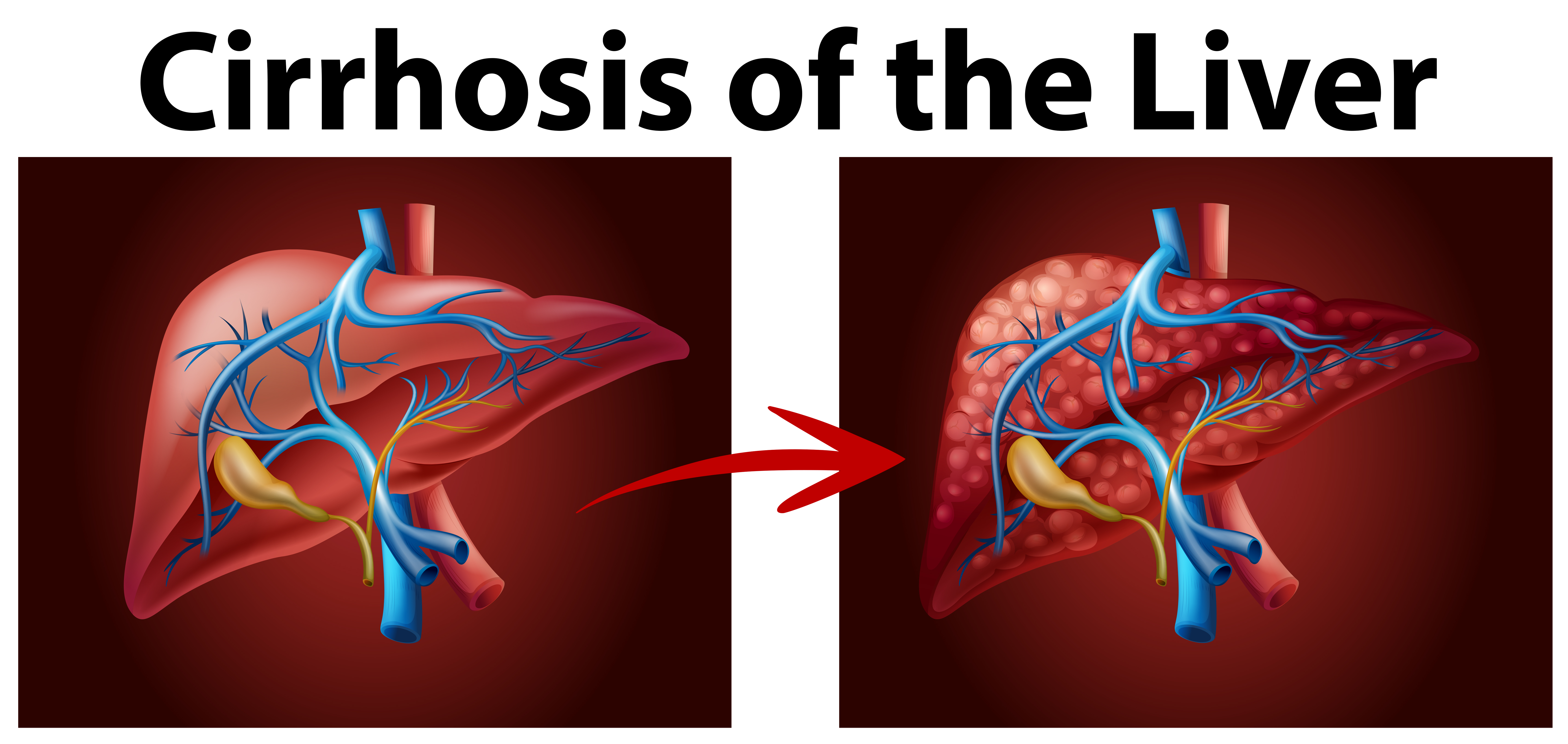
Diagram showing cirrhosis of the liver 292464 - Download ... from static.vecteezy.com The system possesses two steady states of liver volume, k and m. A primary liver cancer is uncommon; The liver is a roughly triangular organ that extends across the entire abdominal cavity just inferior to the diaphragm. Liver medical diagram of body digestive system. The hepatic vascular system is dynamic. Leading out of the liver. Learn vocabulary, terms and more with flashcards, games and other study tools. What is the posterior border of the caudate lobe in couinad's liver segments?
Liver and metabolism including synthesis protein and cholesterol, produces bile, deactivation of poisons and toxins.
708x593 illustration showing the origin of the three hepatic arteries. The liver resides in almost the entire length of the upper abdomen. Functions of the healthy liver. K and m represent saturated volume after liver recovery and shrunken volume after irreversible reduction in size due to liver failure. Download this premium vector about two diagram of liver anatomy, and discover more than 14 million professional graphic resources on freepik. The liver is an organ only found in vertebrates which detoxifies various metabolites, synthesizes proteins and produces biochemicals necessary for digestion and growth. Oxygenated blood from the heart to supply liver cells. While the greatest portion sits in the right hypochondriac region, it extends past the epigastrium and over into the left hypochondriac region. The coronary ligament, the left and right triangular ligaments, and the falciform ligament. The system possesses two steady states of liver volume, k and m. The liver region is further segmented using localized contouring. Leading out of the liver. The diaphragm borders the liver superiorly, laterally, and anteriorly.


0 Komentar